SOUTHERN REGIONAL BEEF COW/CALF HANDBOOK:
DETERMINING PREGNANCY IN CATTLE
ISSUED: 1-78
REVISED:
A.M. Sorensen, Jr. and J.R. Beverly*
This material is made available through assistance from the Kentucky
Beef Cattle Association
* Professor, Department of Animal Science, and Extension animal reproduction
specialist, Texas A&M University.
Economic returns from the beef cattle
industry depend largely on the percent calf crop and the weaning weight
of calves to be sold.
The following discussion describes
a way of improving the calf crop percentage through pregnancy determination
and elimination of non-pregnant cows. This determination, called palpation,
is made by inserting the arm into the rectum and feeling the reproductive
tract for pregnancy indications.
Equipment
Little equipment is needed in palpation.
The individual doing the palpating should wear protective covering on the
arm and hand. This may be a rubber sleeve or a plastic sleeve which covers
the arm to the shoulder. This protection guards against disease and eliminates
irritation of the arm. A lubricant, such as liquid soap, is preferred over
detergent. Use a rubber band to hold the plastic sleeve on the upper arm.
Dry rubber sleeves immediately after use and sprinkle with talcum to avoid
deterioration. Plastic Sleeves may tear after several uses, thereby reducing
protection. Do not attempt to use these later.
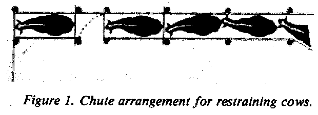
The chute for holding the animal during
palpation should allow the animal to stand on the ground in a normal position.
It should have a front wall or gate and a bar just above the hocks in the
rear, Figure 1. This bar eliminates the animal's
kicking and protects the palpator during manipulation of the reproductive
organs.
Include an entrance gate in the chute
at the rear of the animal to allow entrance and exit for the palpator.
Provide a gate which will swing across the crowding chute in front of other
animals coming behind the palpator. Squeeze chutes may be used. However,
the noise made as the animal enters the chute and her unnatural position
sometimes excite the animal, making palpation more difficult.
Palpation alone takes only a few seconds.
The speed with which pregnancy is determined depends largely on management
of the livestock as they come through the chutes, stage of pregnancy and
the palpator's experience. As many of 800 head of cattle can be palpated
in a normal working day under ideal conditions. However, efficiency is
greatly reduced if the palpator must help bring the cattle into the chute,
climb over the chute wall to get behind the animal and then palpate the
animal.
Palpators should practice certain precautions.
The first of these concerns the palpator's safety. Restrain the animal
so she cannot jump over the side of the chute or kick the palpator. Prevent
other cattle from coming up behind the palpator as he attempts to determine
pregnancy.
Consider also the animal's safety.
Do not place the animal's head in a stanchion or headgate. This tends to
excite the animal. Replace broken boards in the chute that could injure
the animal's legs. A dirt floor chute is most desirable. Animals in a chute
with a slick floor may become excited and lose their footing. Cleats across
the floor stabilize footing.
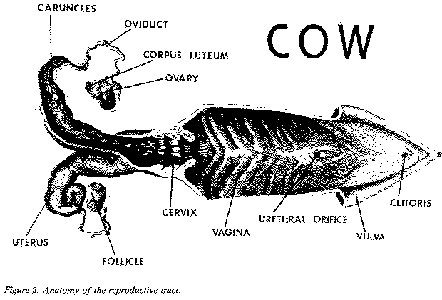
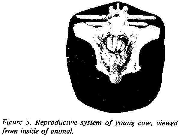
Reproductive System
Thorough knowledge of the female reproductive
system is essential in palpation, Figure 2.
The female germ cell, called the ovum or egg, develops in
a follicle on the ovary. The ovaries are suspended rather
freely in the body cavity by ligaments attached to the top of the
abdominal cavity. These move from one location to another in the cavity.
The ovaries (two) are located on each
side of the cavity. They are approximately 1/2-inch wide, 3/4-inch deep
and l-inch long in a normal cow. This size varies considerably, depending
upon the stage of the estrous cycle.
The ovary should feel firm but not
hard. The follicle which contains the egg is a blister-like projection
on the surface of the ovary. It may reach a size of 1/2 to 3/4 -inch in
diameter and protrude approximately 1/4-inch from the surface. The follicle
has the feel of a blister or tissue filled with fluid. An experienced person
can palpate thc follicle on the ovary.
As the follicle ruptures releasing
the egg, the cavity fills with cells to form another body, called the corpus
luteum,
Figure 3. (Large mature follicle on right
ovary. Mature corpus luteum on left ovary.) This develops as a cellular
mass and protrudes with a teat-like projection at the point of rupture.
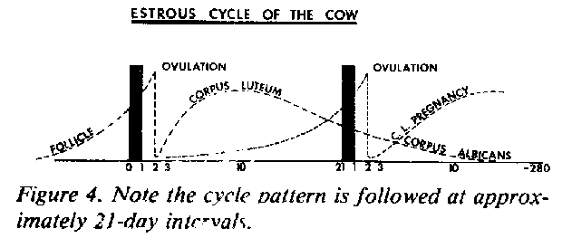
Approximately 15 days after the animal is in estrus, the corpus luteum
begins to regress and almost completely disappears within the next 10 days.
Another follicle is growing and will rupture approximately 21 days after
the previous one. This cycle pattern is followed at approximately 21-day
intervals, Figure 4. The corpus luteum can also
be palpated on the ovary by an experienced person.
The large follicle on the ovary indicates
the animal is approaching the time of estrus. A corpus luteum on the ovary
indicates the animal is in about the midpoint of the estrous cycle or is
pregnant. The corpus luteum persists in the cow throughout pregnancy. Therefore,
palpation of the corpus luteum may either indicate a stage in a normal
cycle or pregnancy.
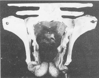
Figure 7. Reproductive tract of mature cow
on the floor of pelvis.
After the egg is released from the follicle,
it moves into the oviduct, a small tube that acts as a passageway
for the egg to go from the ovary into the uterus. The two oviducts
also act as sites of fertilization. The egg normally is fertilized about
one-third of the way down the oviduct by sperm that have entered during
copulation.
The egg moves into the horn of the
uterus and, if fertilization has occurred, begins its cell division. The
egg continues to multiply and lay down its membranes, implanting itself
in one of the uterine horns.
The cow's uterus is made up of two
horns with a connecting body between, figure 2. Therefore, in development
of the membranes, they pass from the tip of one horn through the body to
the tip of the other horn. Attachment takes place throughout. The uterus
is lined with numerous raised prominences, called caruncles, with
form attachment points for developing cotyledons or "buttons" on
the fetal membranes. The next portion of the reproductive tract toward
the exterior is called the cervix, figure 2, and is made up of a
connective tissue substance that feels much like gristle. The cervix is
tortous with folds protruding into the lumen and extending in the direction
of the exterior. Within these folds are numerous glands which secrete fluid
abundantly during estrus. It becomes thick and tenacious during the functioning
period of the corpus luteum and during pregnancy.
The next portion of the tract, the
vagina,
figure 2, acts as a receptacle for the penis during copulation and the
point of deposition of the spermatozoa. The bladder, which opens
on the floor of the vagina through the urethral orifice, from this
point to the vagina exterior acts as a common passageway for urine and
passage of young at birth.
The vagina has the feeling of a thin-walled
organ similar to that of the uterus. The vulva is the external portion
of the reproductive tract and may be seen as two prominent lips.
The entire reproductive organs of an
animal vary considerably in size and feel with the stage of development
during pregnancy and also with the size and reproductive history of the
animal. Generally, the size of the entire nonpregnant reproductive tract
is 12 to 18 inches long. In young heifers that have just reached puberty,
the reproductive organs may be only 8 inches long. The reproductive tract
of older cows that have had several calves may extend to 24 inches. Diameter
of the uterine horns is approximately 3/4 to 1 inch and the length of the
horns 6 to 8 inches with a 3 to 4-inch body. The cervix is approximately
1 to 2 inches in diameter and 3 to 5 inches long.
Developmental Stages
Periods of development in a young calf's
life are divided into three parts. The period of the ovum is that
time from fertilization until the egg has divided enough times to take
on a particular form. This occurs about the thirtieth day when there is
an enfolding of the layers of the developing egg. At this stage, the newly
developing animal is called an embryo. The period of embryonic development
lasts until attachment of the fetal membranes to the lining of the uterus
- approximately 38 days. During the embryonic stage, various organs and
systems are laid down. These include the respiratory system, nervous system,
digestive system, circulatory system and reproductive system.
The embryo, as it develops, floats
freely in the uterine cavity, bathed by a secretion called uterine milk.
During this time, the embryo lays down all of the organs and tissues.
When the embryo is about 38 days old,
the fetus period begins. This term is used until the newborn is
expelled at parturition. During the fetus stage, continued attachment takes
place at the numerous caruncles lining the uterus. These attachments provide
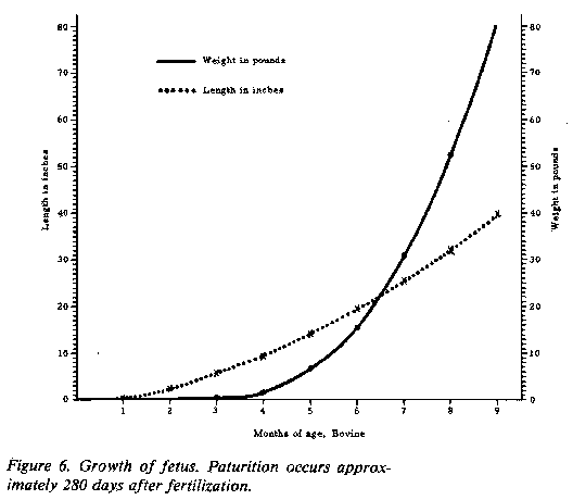
transfer of nutrients and waste materials for the developing fetus. Parturition
occurs approximately 280 days after fertilization, Figure
6.
Palpation
Either hand may be used in palpation.
One hand may grasp the cow's tail as a handle. The other hand should be
well lubricated and shaped into a wedge by bringing the fingers together
as closely as possible. The hand is pushed through the anus into the rectum
with one swift thrust. As the hand enters the rectum, fold the fingers
into a modified fist, Figure 5. By bailing the
hand into a modified fist as it enters the rectum, the fecal material is
pushed aside and the rectum straightened. Folds in the rectum do not straighten
as easily if the fingers are held in a pointed position. This also eliminates
puncturing of the rectal wall with the sharper pointed fingers. However,
puncturing is rare, as the rectum is thick-walled and resistant.
Cleaning the cow's rectum of fecal
material usually is not necessary. However, in early stages of learning,
cleaning the rectum increases feel. Remove fecal material of cattle on
range since it is so dry.
Feeling through the rectal wall is
similar to feeling through a layer or two of thin rubber. Most cattle are
cooperative. Thus, it should be possible to feel the paunch and pick-up
the reproductive organs without difficulty.
Usually, the longer the examination,
the more resistance encountered. Occasionally, a small amount of bleeding
occurs. This should not upset the palpator. An indication of rectum damage
is a sandpaper or gritty feeling. In this case, the mucosa lining the rectum
has been rubbed off in the palpation process. It is best to stop further
palpation when this occurs.
A thrust of the arm to the elbow is
usually much better than trying to put the hand into the rectum and gradually
working forward. It is much easier to work to the rear, since that is the
direction the cow is pushing the fecal matter and the inserted arm. In
palpating, assume the animals are pregnant. Therefore, reach farther than
wrist deep to pick up the uterus and the calf within.
Certain landmarks are evident inside
the cow. The pelvis forms a bone cradle for the reproductive system,
Figure 5. The nonpregnant tract usually is located near the top of the
pelvic cradle and felt easily with downward pressure. As pregnancy advances,
the uterus and cervix move down and into the body cavity.
The cervix with its firm feel
is also a good landmark, figure 2. After the locating the cervix, the palpator
can move forward to the uterus to determine pregnancy.
The paunch, located directly forward
and to the left, may feel like the end of a football and be rather soft
and mushy. The feel depends on the amount of feed in the paunch. The feedstuff
when mashed slowly returns to normal shape. It does not have the
watery, soft feel of the pregnant uterus.
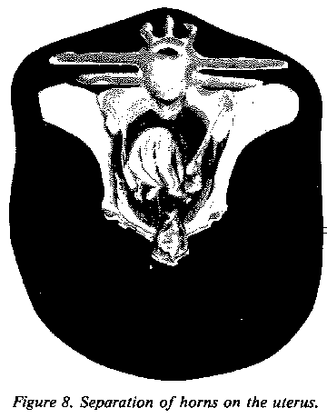
The "open" reproductive tract normally
lies on the floor of the pelvis. The horns of the uterus are coiled on
the front edge of the pelvis or, in older cows, may hang slightly into
the abdominal cavity. The entire tract may be held in the hand at this
stage. Slight pressure by the middle finger will separate the horns of
the uterus, Figure 8. The ovaries are located
in the broad ligament on each side.
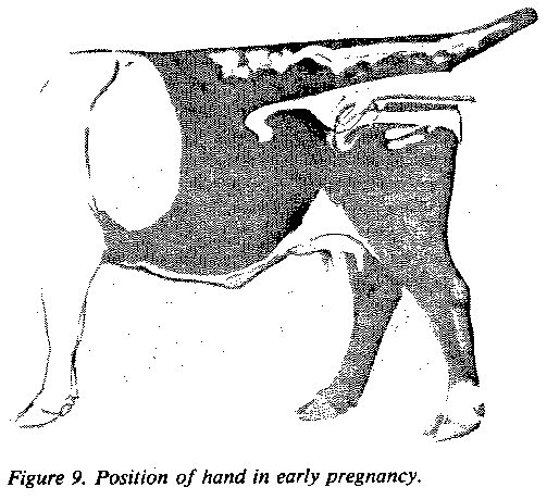
30-day pregnancy. A palpator,
with skill and practice, can detect pregnancy as early as 30 days after
breeding. Palpation at this early stage should be accompanied by good breeding
herd records. The palpator through these records knows the approximate
breeding date of the animal.
In the early stage of pregnancy, the
uterus, filled with a small amount of fluid, will feel slightly thinner.
One horn is enlarged a little more than the other. Presence of the embryonic
vesicle at this time is determined by running the horn between the fingers
in a milking action to feel the vesicle pop through the fingers, Figure
9.
The embryo is only about 1/2-inch long.
However, the vesicle surrounding it is approximately 3/4-inch in diameter
and filled with fluid, such as a balloon filled tightly with water. On
the same side as the enlargement, the palpator will find a corpus luteum
on the ovary. The uterus, in much the same location as a nonpregnant uterus,
has not been displaced because of size or weight at this time. The outer
embryonic vesicle, which is rather thin with little fluid, may be 18 to
24 inches long. By pinching the horn of the uterus carefully, the membranes
of this vesicle are felt as they slip between the fingers.
Figure 10. 60-day pregnancy. Uterus hangs
over pelvic brim..
45-day pregnancy. Most palpators
prefer bulls be separated from cows at least 45 days before pregnancy determination.
At 45 days, one horn of the uterus containing the fetus is somewhat enlarged
and thinner wailed and the corpus luteum is on the ovary of the same side.
The fetus at this stage is approximately 1 inch long. The vesicle around
it is somewhat egg-shaped and measures approximately 1 to 1 1/2 inches
long. The outer membrane, which contains considerable fluid, may be felt
through the uterine wall. Attachment of the membranes to the uterus has
just taken place at approximately 38 to 40 days. Therefore, avoid moving
the fetus about in the uterus. The caruncles on the uterus join the cotyledons
on the fetal membranes for nutrient exchange.
60-day pregnancy. The uterus
has enlarged until one horn is about the size of a banana, measuring 8
to 10 inches long. Weight of the contents pulls the uterus into the body
cavity just over the pelvic brim, Figure 10. The fetus has grown rapidly
and, at this stage, is about 2 1/2 inches long. The embryonic vesicles
are still prominent and, at this stage, may be felt without feeling the
fetus.
The uterine walls have thinned considerably.
The best method of feeling the fetus is to bobble it with your hand so
that by gently tapping the uterus the fetus swings as a pendulum and hits
against the wall of the uterus and vesicle. The cervix remains on top of
the pelvic cradle with the uterine horns moving forward and downward over
the brim. The ovaries are still suspended by the broad ligaments and in
early stages will be rather high in relation to the uterus. As before,
a corpus luteum should be on the ovary of the same side as the developing
fetus. The presence of the fetus eliminates a need for feeling other structures.
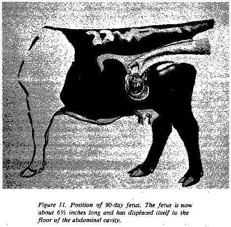
90-day pregnancy. The uterus
will have enlarged considerably by this time, filled with fluid and increased,
growth of the fetus, Figure 11. The fetus now
is about 6 1/2 inches long and has displaced itself to the floor of the
abdominal cavity, indicating the uterus has stretched considerably. The
cervix may be pulled to the pelvic brim so that the cervix, body and horns
of the uterus are in the abdominal cavity.
The ovaries are usually pulled down
with the uterus to much lower than normal and may be palpated to either
side of the uterus. In larger animals, this is a difficult stage of pregnancy
because of displacement and the distance from the anus to the developing
fetus.
Factors other than presence of the
fetus itself may have to be considered at this stage. Displacement of the
uterus, an indication of pregnancy, should be considered. Another indication
of pregnancy is enlargement of the uterine artery with its characteristic
"whirring': pulsation. This artery passes in the forward fold of the broad
ligament supporting the uterus. At 3 months, it is approximately 1/8 to
3/16 inch in diameter. The pulse of the heart beat is felt easily as blood
is carried into the uterus to nourish the developing fetus.
Do not confuse the uterine artery with
the femoral artery lying on the inside of the thigh which supplies the
hind legs. The femoral artery is lying in the muscle but may be palpared.
Remember that the uterine artery is in the broad ligament and may be moved
4 to 6 inches, whereas the femoral may not. Another pregnancy indication
is presence of a corpus luteum on one of the ovaries, although this corpus
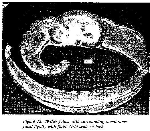
luteum may appear here even in a normal cycle. The best indication of pregnancy
in absence of the fetus is the presence of cotyledons. Cotyledons in a
3-month pregnancy should be flattened and egg-shape and measure 3/4 to
l-inch across. Although rather soft to the touch, they are firmer than
the thin-walled uterus. The membranes still are filled tightly with fluid,
Figure
12.
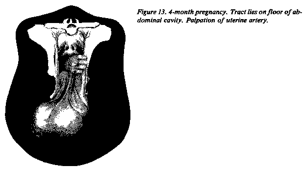
120-day pregnancy. At this stage,
the fetus is displaced similarly to the 90-day fetus. However, it has enlarged
to approximately 10 to 12 inches long. The head is about the size of a
lemon. Often the head of the developing fetus is picked up before any other
part.
The enlarged fetus fills a greater
portion of the abdominal cavity and is easier to feel than the 3-month
fetus, Figure 13. All other characteristics
have changed some. Presence of the cotyledons is more noticeable, since
they have developed to approximately 1 1/2 inches in length. The pulsating
uterine artery may be palpated, as well as the corpus luteum and displacement
of the entire reproductive tract.
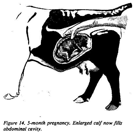
Over 5-month pregnancy. The
main change until parturition will be in size, Figure
14, as the fetus enlarges rapidly utilizing more of the abdominal cavity.
Table 1 summarizes outstanding identifying characteristics.
Other Factors
The paunch.As one reaches into
the rectum, feeling directly forward and to the left, the dorsal posterior
sac of the paunch may be palpated. This paunch in an animal on good pasture
or on full feed will be rather firm and plastic to the touch. By mashing
the paunch you notice an indentation which gradually smooths back over
indicating that the paunch is full of feedstuff. This dorsal posterior
sac may feel much like the end of a football, coming to somewhat of a point.
This may be misinterpreted under careless examination as a large uterus
in latter stages of pregnancy.
TABLE 1. FETAL SIZE AND CHARACTERISTICS USED IN DETERMINING PREGNANCY
| Days of Gestation |
Fetal Size |
Identifying Characteristics |
| Weight |
Length (Inches) |
| 30 |
1/100 oz. |
2/5 |
One uterine horn slightly enlarged and thin; embryonic vesicle size
of large marble. Uterus in appoximate position of nonpregnant uterus. Fetal
membranes may be slipped between fingers from 30 to 90 days. |
| 45 |
1/8-1/4 oz. |
1-1 1/4 |
Uterine horn somewhat enlarged, thinner walled and prominent. Embryonic
vesicle size of hen's egg. |
| 60 |
1/4-1/2 oz |
2 1/2 |
Uterine horn size of banana; fluid filled and pulled over pelvic brim
into body cavity. Fetus size of mouse. |
| 90 |
3-6 oz. |
5-6 |
Both uterine horns swollen (3 to 3 " in diameter) and pulled deeply
into body cavity (difficult to palpate). Fetus is size of rat. Uterine
artery 1/8 to 3/16" in diameter. Cotyledons 3/4 to 1" across. |
| 120 |
1-2 lb. |
10-12 |
Similar to 90-day but fetus more easily palpated. Fetus is size of
small cat with head the size of a lemon. Uterine artery 1/4" in diameter.
Cotyledons more noticeable and 1 1/2 inches in length. Horns are 4 to 6"
in diameter. |
| 150 |
4-6 lb. |
12-16 |
Difficult to palpate fetus. Uterine horns are deep in body cavity with
fetus size of large cat--horns 6-8" in diameter. Uterine artery 1/4-3/8
in diameter. Cotyledons 2 to 2" in diameter. |
| 180 |
10-16 lb. |
20-24 |
Horns with fetus still out of reach. Fetus size of small dog. Uterine
artery 3/8-1/2" in diameter. Cotyledons more enlarged. From sixth month
until calving a movement of fetus may be elicited by grasping the feet,
legs or nose. |
| 210 |
20-30 lb. |
24-32 |
From 7 months until parturition fetus may be felt. Age
is largely determined by increase in fetal size. The uterine artery continues
to increase in size--210 days, 1/2" in diameter, 240 days.to 5/8" in diameter;
270 days, 1/2 to 3/4" in diameter. |
| 240 |
40-60 lb. |
28-36 |
| 270 |
60-100 lb. |
28-38 |
Cotyledons. Cotyledons may be
interpreted as ovaries or vice versa. Cotyledons do not have the solid
feel of an ovary but are rather soft. The best comparison is to that of
dried apricots soaked in water. The ovaries are more rounded and egg-shaped
with a firm feel. Only two are present.
Pyometra. In this condition,
the uterus is filled with white blood cells attempting to clear up disease
organisms. The uterus may be fluid to the touch or may be somewhat solidified,
feeling rather plastic. This stage may be confused with early pregnancy
stages if the uterus is in a fluid condition and only partly filled. In
the latter stages of pyometra, the uterus becomes rather firm.
Large uteri. In older cows that
have had many calves, the uterus may not return to its normal size as in
a younger cow. The enlarged uterus may feel as if displaced over the brim
of the pelvis as in a 3 to 4-month pregnancy. Careful manipulation of the
uterus shows no fluid and no cotyledons developing in the open cow. Relaxation
of the broad ligament tends to cause a similar condition.
Bladder. The urinary bladder
may be interpreted as pregnancy in the 60 to 75-day stages. At this time,
the full bladder feels similar to the uterus filled with fluid. Careful
tracing should indicate a bladder, where there is only one body, or a pregnant
horn of the uterus, where both horns can be palpated and traced back to
the cervix.
Enlarged cervix. In some Brahman
and Brahman crossbred cattle, an enlarged cervix is found that is firm
and has the feel of a developing fetus in the latter stages. Tracing the
reproductive tract distinguishes between the two.
Breed differences. Brahman,
Brahman crossbred, Santa Gertrudis, Charolais, Holstein and Brown Swiss
cattle, because of their increased size, are slightly more difficult to
palpate in certain stages of pregnancy than the smaller European breeds.
In 3 to 4-month stages, the uterus
has dropped so deeply into the body cavity it is almost impossible to palpate.
In such instances, pass the hand under the cervix and lift the uterus to
feel the fetus itself. By lifting the uterus and quickly moving the hand
down into the body cavity, the presence of the fetus is felt by bobbing
the fluid and the fetus through the wall of the uterus.
Brahman and Charolais breeds appear
to have more tissue inside than smaller breeds. More folds of the omentum
seem to cover the intestines, making it slightly more difficult to pick
up the uterus.
Charolais cattle seem to have less
flexibility in the rectum. It is commonly harder to feel deep in the body
cavity in these cattle, and lateral movement is somewhat restricted.
The uteri of heifers of Brahman breeding
vary considerably. It is not uncommon to find l,000-pound heifers with
uteri measuring only 4 to 6 inches in length, as compared to a normal uterus
which would be 10 to 12 inches.
Highly finished cattle for show or
on lush pastures may be filled with fat which interferes with movement
and feel. These cattle are very difficult to palpate. Repalpate at a later
date in case of doubt.
Recommendations
Practice! Experience is the key to
palpation. In many instances the ranch manager should not be the one to
palpate but should supervise the operation and critically observe the cows.
Unhealthy, unsound and undesirable types should be eliminated as well as
open cows.
Shorten the calving interval by reducing
the time during the breeding season when the bulls are with the cows. Cows
that settle first are those most adapted to reproduction. Wait approximately
45 days after the bulls are removed to palpate. Most cows should conceive
at the beginning of the season, and only a few will be short-tern pregnancies.
Cull as critically as feasible. If
every open, unsound cow can be removed, cull immediately.
Remember, palpation is an art and a
skill. It pays dividends to the person who uses it wisely.